Advanced Solutions
for Advanced Pathology
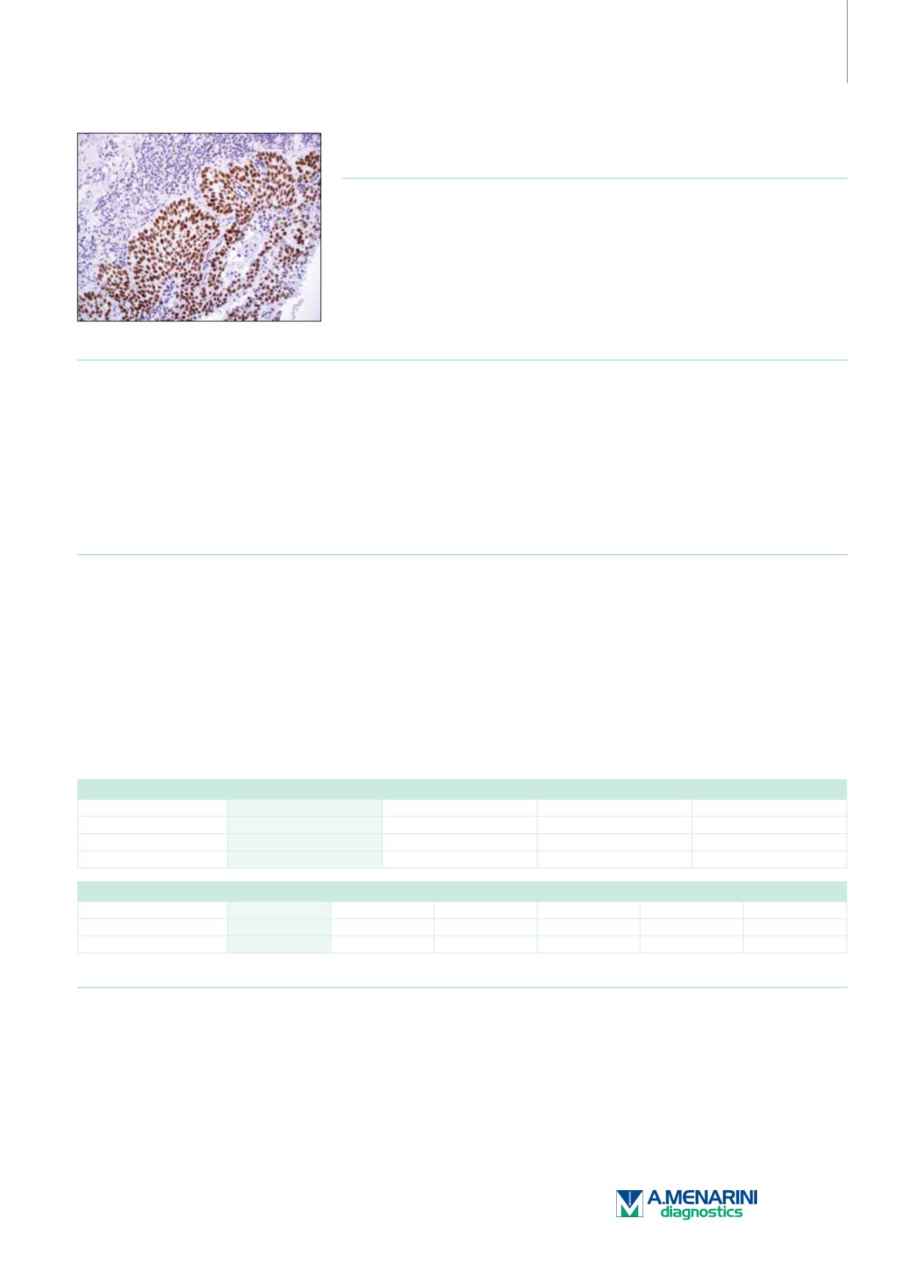
SOX-2 (SP76)
Rabbit Monoclonal Antibody
Cat. No. Description
Volume
45334 IMPATH SOX-2 RTU R (SP76)
50 Tests
44389 SOX-2 RTU R (SP76)
7 ml Ready To Use
44797 SOX-2 0,1 R (SP76)
100 µl liquid Concentrated
44798 SOX-2 1 R (SP76)
1 ml liquid Concentrated
Product Specifications
Designation
IVD
Reactivity
Paraffin
Visualization
Nuclear
Control
Lung Squamous Carcinoma
Stability
Up to 36 mo. at 2-8°C
Isotype
IgG
Manual Protocol*
• Pretreatment: Heat Induced Epitope
Retrieval (HIER)
• Primary Antibody Incubation Time:
10-30min @ 25-37°C
• 2-step polymer detection
*Please refer to product insert for complete protocol.
ImPath Protocol*
• Dewax: Dewax Solution 2 (DS2)
• Pretreatment: Retrieval Solution pH 9.0
(TR1) 32min @ 98-103°C
• Primary Antibody Incubation Time:
10-90min @ 25-37°C
• HRP Polymer (Universal) or AP Polymer
(Universal) for 12 min
*Please refer to product insert for complete protocol.
Product Description
Anti-SOX-2 recognizes lung squamous cell carcinoma (LSCC). Extensive anti-SOX-2 staining is seen in over 90% of LSCC and largely parallels
p63 expression. However, only 4.5% of lung adenocarcinoma (LACA) is positive for SOX-2. In a study by Sholl et al. 29% of LACA cases
exhibited at least focal p63 expression. Combined p63 and SOX-2 expression was seen in 94% of LSCC and 12% of LACA with a statistically
significant difference (P<0.0001) versus p63 alone. Anti-CK 5&6 had a good sensitivity but poor specificity for LSCC. Combined anti-CK 5&6 and
anti-p63 positivity was seen in 93% of LSCC and 24% of LACA. Anti-CK 5&6+/anti-p63+/anti-SOX-2+ was detected in 93% of LSCC and only
9% of LACA. These results indicate that the sensitivity of anti-p63 is equally high but its specificity is similarly variable; it was seen at least focally
in close to 30% of LACA. When used together, anti-p63+/anti-SOX-2+ applied to the same tumor cell population is >90% specific for LSCC.
Anti-SOX-2 produced moderate-to-intense staining in all 50 cases of embryonal carcinoma components with strong anti-SOX-2 positivity and
moderate-to-intense staining. The only other component that showed reactivity was the primitive neuroectodermal component in 11 of 14 (79%)
of immature teratomas. In each of these positive staining foci, the staining varied from moderate-to-strong. Yolk sac tumor, seminoma, mature
teratoma, choriocarcinoma, and IGCNU were uniformly negative, as were all the non-neoplastic parenchymal and stromal structures.
Lung
SOX-2
p63
Napsin A
TTF-1
Lung Adenocarcinoma
-
-/+
+
+
Lung SQ Carcinoma
+
+
-
-
Lung NET
-/+
-
-
+
Germ Cell Tumor
SOX-2
Oct-4
SALL4
CD117
CD30
PLAP
Seminoma
-
+
+
+
-
+
Embryonal Carcinoma
+
+
+
-
+
-
Reference
1. Sholl LM, et al. Appl Immunohistochem Mol Morphol. 2010; 18:55–61.
2. Tsuta K, et al. J Thorac Oncol. 2011; 6:1190– 1199.
3. Gopalan A, et al. Mod Pathol. 2009; 22:1066–1074.
217


