Advanced Solutions
for Advanced Pathology
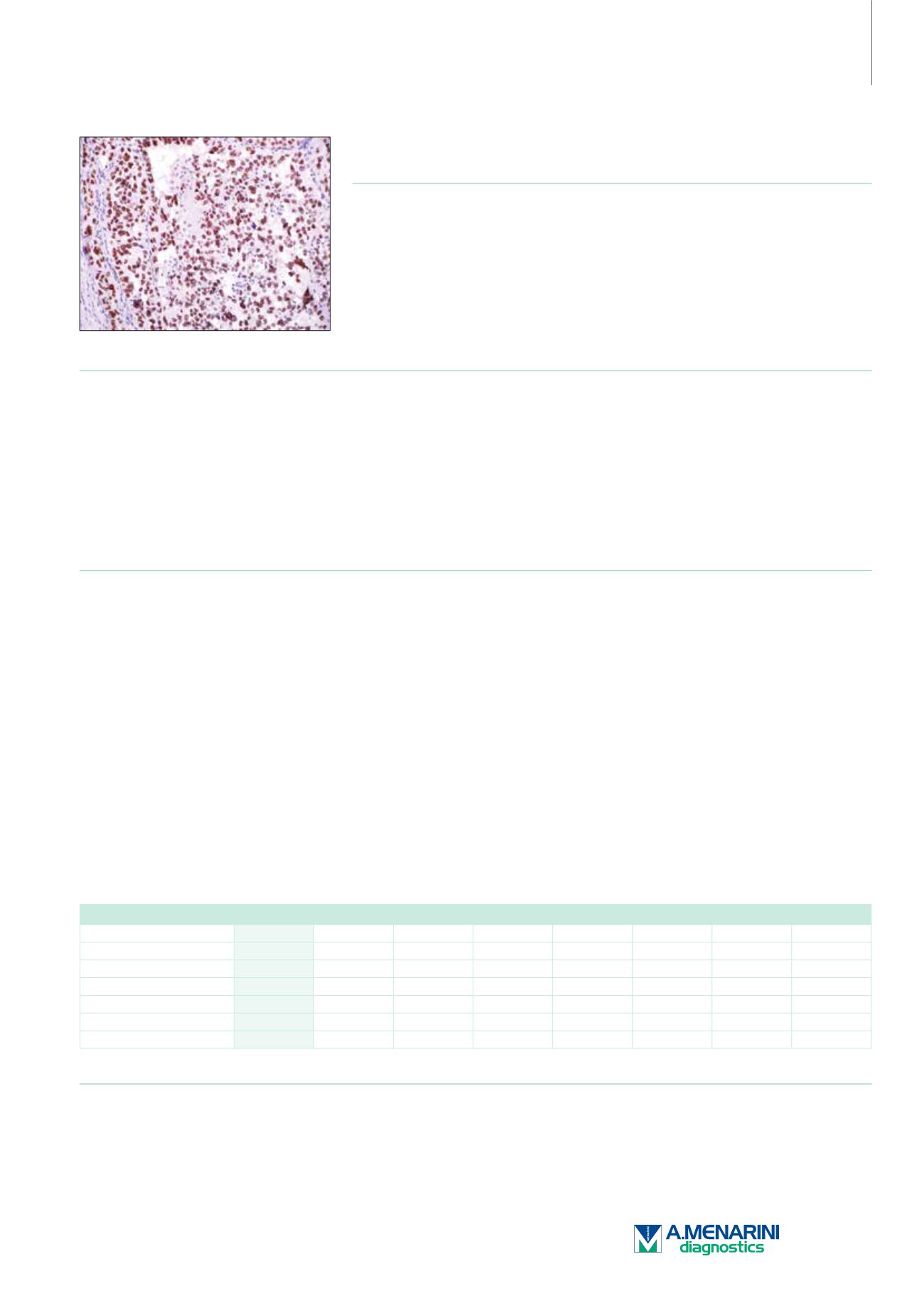
TFE3 (MRQ-37)
Rabbit Monoclonal Antibody
Cat. No. Description
Volume
45339 IMPATH TFE3 RTU R (MRQ-37)
50 Tests
44397 TFE3 RTU R (MRQ-37)
7 ml Ready To Use
44813 TFE3 0,1 R (MRQ-37)
100 µl liquid Concentrated
44814 TFE3 1 R (MRQ-37)
1 ml liquid Concentrated
Product Specifications
Designation
IVD
Reactivity
Paraffin
Visualization
Nuclear
Control
Melanoma, Testis, Xp11.2
translocation renal cell carcinoma
Stability
Up to 36 mo. at 2-8°C
Isotype
IgG
Manual Protocol*
• Pretreatment: Heat Induced Epitope
Retrieval (HIER)
• Primary Antibody Incubation Time:
10-30min @ 25-37°C
• 2-step polymer detection
*Please refer to product insert for complete protocol.
ImPath Protocol*
• Dewax: Dewax Solution 2 (DS2)
• Pretreatment: Retrieval Solution pH 9.0
(TR1) 32min @ 98-103°C
• Primary Antibody Incubation Time:
10-90min @ 25-37°C
• HRP Polymer (Universal) or AP Polymer
(Universal) for 12 min
*Please refer to product insert for complete protocol.
Product Description
Xp11 translocation renal cell carcinoma (RCC) is a recently recognized subset of RCC, characterized by chromosome translocations involving
the Xp11.2 break point and resulting in gene fusions involving the TFE3 transcription factor gene that maps to this locus. Xp11 translocation
RCC represents the most common type of RCC in children, but is less frequent on a percentage basis in adults. Morphologically, the neoplasm
frequently shows papillary architecture and clear cytoplasm, and frequently has associated psammoma bodies. Immunohistochemically, the
neoplasm under-expresses epithelial markers such as cytokeratin and epithelial membrane antigen compared with typical RCC. The most
sensitive and specific immunohistochemical marker for the Xp11 translocation RCC is nuclear labeling of TFE3 protein, which reflects over-
expression of the resulting fusion proteins relative to TFE3. Alveolar soft part sarcoma (ASPS) is an uncommon soft tissue sarcoma which affects
predominantly young patients, often in the extremities.
ASPS has the specific molecular translocation der(17)t(X;17)(p11.2;q25), which fuses the TFE3 transcription factor gene at 17q25 to ASPL,
a gene at 17q25 to form a fusion transcript of ASPL-TFE3. The diagnosis of ASPS can be problematic due to histologic overlap with other
tumors, particularly in small biopsies, as well as when the detection of a metastasis is prior to identification of a primary, or when presenting
at unusual primary sites such as bone. Moreover, there has previously been a lack of specific diagnostic markers. The differential diagnoses
include, in particular, paraganglioma, granular cell tumor, metastatic renal cell carcinoma, hepatocellular carcinoma, melanoma, and adrenal
cortical carcinoma. Carcinomas can be separated by the expression of cytokeratins. Paraganglioma shows very strong positivity with
anti-synaptophysin. Melanomas can be distinguished by strong positivity with antibodies against HMB-45, S100, and Melan A. These markers
generally are all negative in ASPS. Anti-TFE3 has been shown to be highly specific and sensitive for ASPS.
Carcinomas
TFE3
RCC
CD10
CK 7 Ksp-cadherin S100P
CD117
CK, HMW
Xp11 Tr RCC
+
+
+
-/+
+
-
Clear Cell RCC
-
+
+
-/+
-/+
-
-
-
Papillary RCC
-
+
+
+
-/+
-
-
+/-
Chromophobe RCC
-
+
+/-
+
+
-
+
-
Oncocytoma
-
-
+
-/+
+
-
+
-/+
Urothelial Carcinoma
-
-
+
+
-
+
+/-
+/-
Reference
1. Argani P. Am J Clin Pathol. 2006; 126(3):332–334.
2. Argani P, et al. Am J SurgPathol. 2003; 27(6):750–761.
3. Argani P, et al. Clin Lab Med. 2005; 25(2):363–378.
4. Lazar AJ, et al. Histopatholo. 2009; 55:750–755.
227


