Antibodies for
Immunohistochemistry
p57
Kip2
(Kp10)
Mouse Monoclonal Antibody
Cat. No. Description
Volume
45248 IMPATH P57 RTU M (Kp10)
50 Tests
44362 p57 RTU M (Kp10)
7 ml Ready To Use
44744 p57 0,1 M (Kp10)
100 µl liquid Concentrated
44745 p57 1 M (Kp10)
1 ml liquid Concentrated
Product Specifications
Designation
IVD
Reactivity
Paraffin
Visualization
Nuclear
Control
Placenta
Stability
Up to 36 mo. at 2-8°C
Isotype
IgG
2b
/k
Manual Protocol*
• Pretreatment: Heat Induced Epitope
Retrieval (HIER)
• Primary Antibody Incubation Time:
10-30min @ 25-37°C
• 2-step polymer detection
*Please refer to product insert for complete protocol.
ImPath Protocol*
• Dewax: Dewax Solution 2 (DS2)
• Pretreatment: Retrieval Solution pH 9.0
(TR1) 32min @ 98-103°C
• Primary Antibody Incubation Time:
10-90min @ 25-37°C
• HRP Polymer (Universal) for 12 min
*Please refer to product insert for complete protocol.
Product Description
p57KIP2 is a cyclin-dependent kinase inhibitor, cell cycle inhibitor and tumor suppressor gene, located at 11p15.5. p57KIP2 shows strong
paternal genomic imprinting, resulting in expression predominantly from the maternal allele.
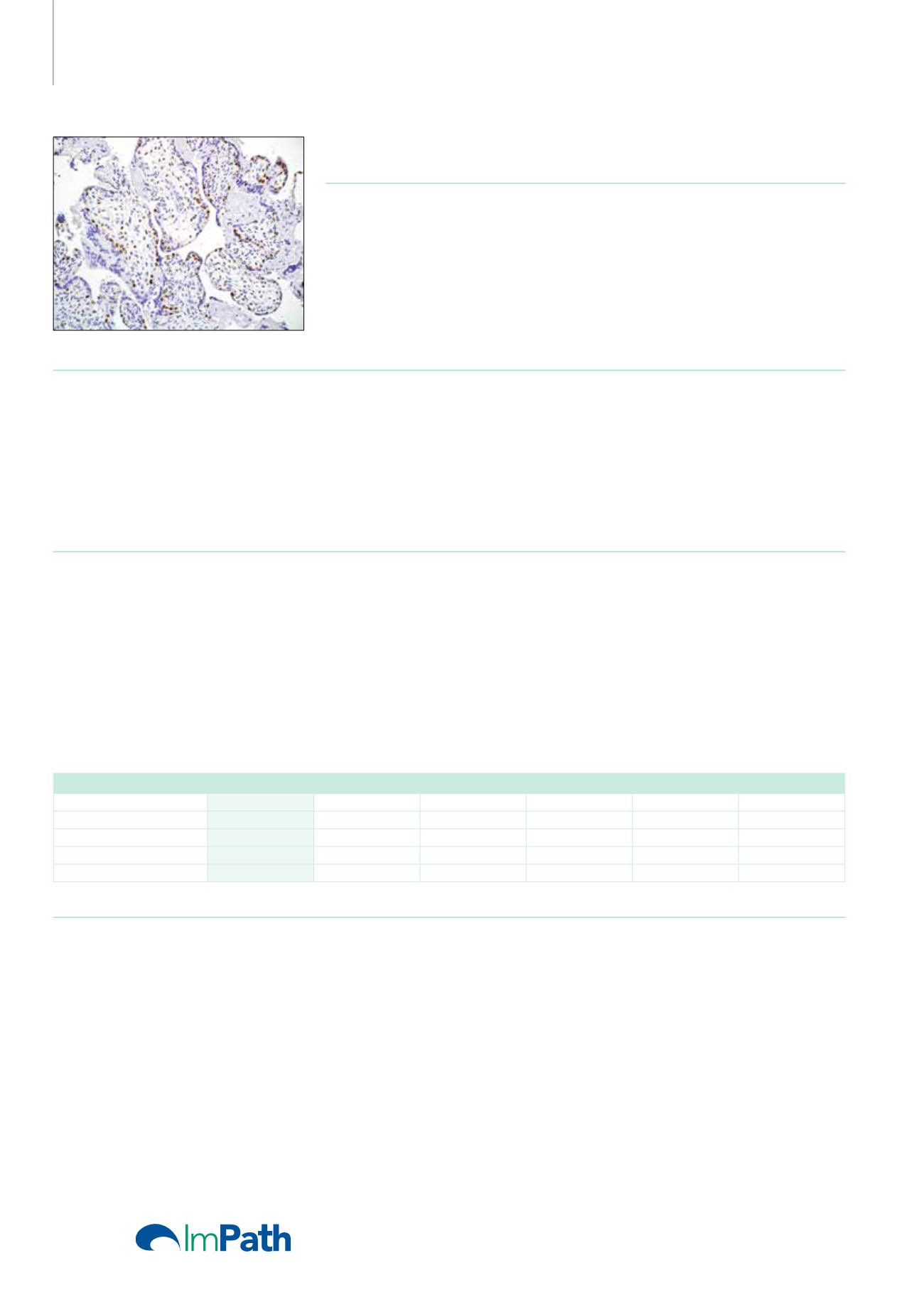
Anti-p57 has been used as an aide in identification of complete hydatidiform mole (CHM) (no nuclear labeling of cytotrophoblasts and stromal
cells) from partial hydatidiform mole (PHM) in which both cytotrophoblasts and stromal cells stain. The histological differentiation of complete
mole, partial mole, and hydropic spontaneous abortion is problematic. Most complete hydatidiform moles are diploid, whereas most partial
moles are triploid. Ploidy studies will identify partial moles, but will not differentiate complete moles from non-molar gestations. Complete moles
carry a high risk of persistent disease and choriocarcinoma, while partial moles have a very low risk. In normal placenta, many cytotrophoblast
nuclei and stromal cells are labeled with this antibody. Similar findings apply to PHM and hydropic abortus tissues. Intervillous trophoblastic
islands (IVTIs) demonstrate nuclear labeling in all three entities and serve as an internal control. Other markers which may be useful in a panel for
differentiating the various forms of gestational trophoblastic disease are anti-hCG, anti-placental alkaline phosphatase, and anti-hPL.
Uterus: Trophoblastic Proliferations
p57
hCG
PLAP
hPL
CK Cocktail
Vimentin
Partial Mole
+
Weak, diffuse
+
Weak, diffuse
Strong, diffuse
-
Complete Mole
-
Strong, diffuse
Weak, focal
Weak, focal
Strong, diffuse
-
Choriocarcinoma
-
Strong, diffuse
Weak, focal
Weak, focal
Strong, diffuse
-/+
Placental Site Tumor
Strong, focal
Strong, diffuse Strong, diffuse Strong, diffuse Strong, diffuse
Reference
1. Kihara M, et al. J Reprod Med. 2005 May; 50(5):307-12.
2. Romaguera RL, et al. Fetal PediatrPathol. 2004 Mar-Jun; 23(2-3):181-90.
3. Marjoniemi VM. Pathology. 2004 Apr; 36(2):109-19. Review.
4. Jun SY, et al. Histopathology. 2003 Jul; 43(1):17-25.
190


