Antibodies for
Immunohistochemistry
Arginase-1 (SP156)
Rabbit Monoclonal Antibody
Cat. No. Description
Volume
45291 IMPATH Arginase-1 RTU R (SP156)
50 Tests
44201 Arginase-1 RTU R (SP156)
7 ml Ready To Use
44429 Arginase-1 0,1 R (SP156)
100 µl liquid Concentrated
44430 Arginase-1 1 R (SP156)
1 ml liquid Concentrated
Product Specifications
Designation
IVD
Reactivity
Paraffin
Visualization
Cytoplasmic, Nuclear
Control
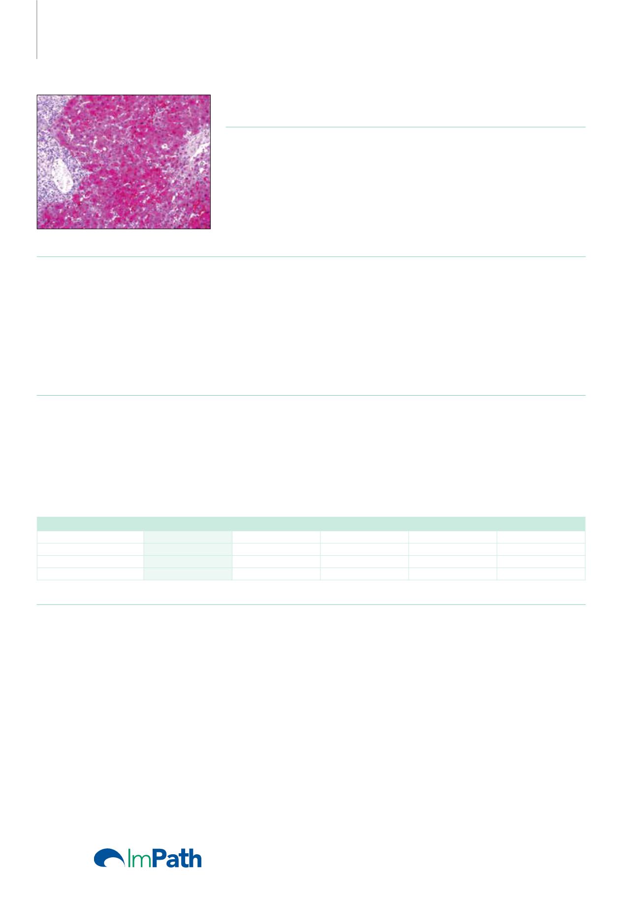
Hepatocellular Carcinoma, Normal
Liver
Stability
Up to 36 mo. at 2-8°C
Isotype
IgG
Manual Protocol*
• Pretreatment: Heat Induced Epitope
Retrieval (HIER)
• Primary Antibody Incubation Time:
10-30min @ 25-37°C
• 2-step polymer detection
*Please refer to product insert for complete protocol.
ImPath Protocol*
• Dewax: Dewax Solution 2 (DS2)
• Pretreatment: Retrieval Solution pH 9.0
(TR1) 32min @ 98-103°C
• Primary Antibody Incubation Time:
10-90min @ 25-37°C
• HRP Polymer (Universal) or AP Polymer
(Universal) for 12 min
*Please refer to product insert for complete protocol.
Product Description
Hepatocellular carcinoma (HCC) is the most common primary malignant tumor of the liver accounting for an estimated 70% -85% of total
liver cancers worldwide. Diagnostic pitfalls exist in the morphologic distinction of HCC from other hepatocellular and non-hepatocellular
mass lesions. In difficult or equivocal cases, the application of immunohistochemical (IHC) panels has been shown to aid in the distinction of
benign and malignant liver lesions. In particular, the application of IHC using antibodies against CD10, polyclonal carcinoembryonic antigen,
alpha-fetoprotein, HepPar-1, and glypican-3 (GPC-3) has proven valuable in liver biopsy and FNA cytology specimens. Recent studies have
shown the usefulness of anti-Arginase-1 as an IHC marker of hepatocellular differentiation in benign and malignant lesions of liver on both
biopsy as well as fine needle aspiration specimens. Arginase-1 expression was present in all (100%) of well-differentiated HCC, 92% cases of
moderately differentiated HCC and was absent in all cases of poorly differentiated HCC (0%).
Liver Neoplasms
Arginase-1
Hep Par-1
Glypican-3
CD10
pCEA
Hepatic Adenoma
+
+
-
+
+
Hepatocellular Carcinoma
+
+
+
+
+
Metastatic Adenocarcinoma
-
-
-
-/+
-/+
Reference
1. Wee A. Cytopathology. 2011; 22:287-305.
2. Nassar A, et al. Diagnostic Cytopathology. 2009; 37:629-635.
3. Yan BC, et al. Am J SurgPathol. 2010; 34:1147-1154.
28


