Advanced Solutions
for Advanced Pathology
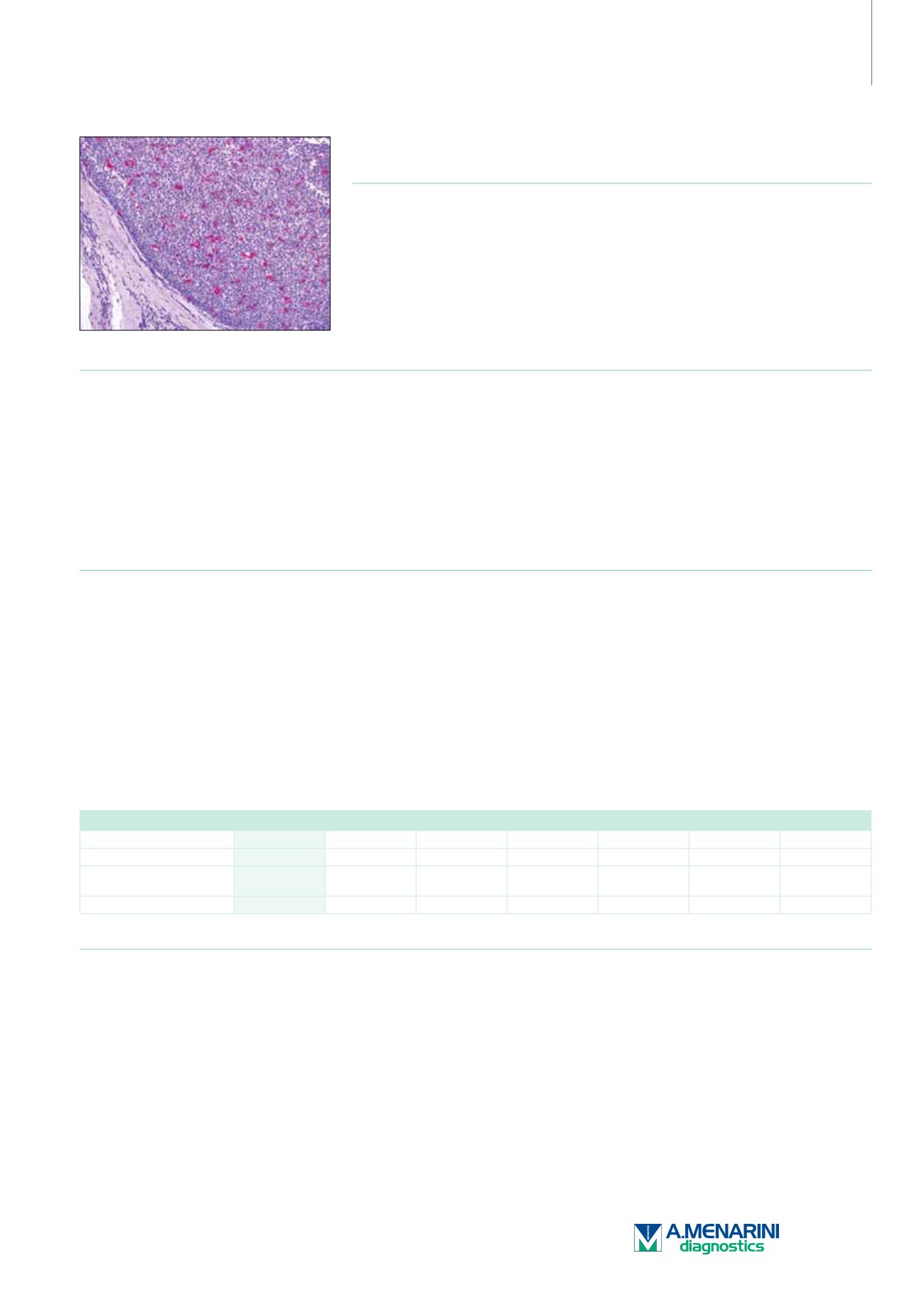
Macrophage (HAM-56)
Mouse Monoclonal Antibody
Cat. No. Description
Volume
45225 IMPATH Macrophage RTU M (HAM-56)
50 Tests
44330 Macrophage Marker RTU M (HAM-56)
7 ml Ready To Use
44682 Macrophage Marker 0,1 M (HAM-56)
100 µl liquid Concentrated
44683 Macrophage Marker 1 M (HAM-56)
1 ml liquid Concentrated
Product Specifications
Designation
IVD
Reactivity
Paraffin
Visualization
Cytoplasmic
Control
Tonsil
Stability
Up to 36 mo. at 2-8°C
Isotype
IgM/k
Manual Protocol*
• Pretreatment: Heat Induced Epitope
Retrieval (HIER)
• Primary Antibody Incubation Time:
10-30min @ 25-37°C
• 2-step polymer detection
*Please refer to product insert for complete protocol.
ImPath Protocol*
• Dewax: Dewax Solution 2 (DS2)
• Pretreatment: Retrieval Solution pH 9.0
(TR1) 32min @ 98-103°C
• Primary Antibody Incubation Time:
10-90min @ 25-37°C
• HRP Polymer (Universal) or AP Polymer
(Universal) for 12 min
*Please refer to product insert for complete protocol.
Product Description
Macrophages comprise many forms of mononuclear phagocytes found in tissues. Mononuclear phagocytes arise from hematopoietic stem
cells in the bone marrow. After passing through the monoblast and promonocyte states to the monocyte stage, they enter the blood, where
they circulate for about 40 hours. They then enter tissues and increase in size, phagocytic activity, and lysosomal enzyme content becoming
macrophages. When a monocyte enters damaged tissue through the endothelium of a blood vessel (known as the leukocyte extravasation),
it undergoes a series of changes to become a macrophage. Monocytes are attracted to a damaged site by chemical substances through
chemotaxis, triggered by a range of stimuli including damaged cells, pathogens, and cytokines released by macrophages already at the site.
At some sites such as the testis, macrophages have been shown to populate the organ through proliferation. Unlike short-lived neutrophils,
macrophages survive longer in the body up to a maximum of several months.
Anti-HAM-56 reacts with tingible macrophages, interdigitating macrophages of lymph nodes, and tissue macrophages, e.g. Kupffer cells of
the liver and alveolar macrophages of the lung. The antibody also stains a subpopulation of endothelial cells, most prominently those of the
capillaries and smaller blood vessels. Anti-HAM-56 reacts with monocytes, but is unreactive with B- and T-lymphocytes.
Histiocytic Proliferation
HAM-56
S-100
CD68
Vimentin
Lysozyme
CD1a
Factor XIIIa
Juvenile Xanthogranuloma
+
-
+
+
+
-
+
Langerhans Cell
Histiocytosis
+
+
+
+
+
+
-
Dermatofibroma
-
-
+
+
-
-
+
Reference
1. Gown AM, et al. Am J Pathol. 125:191-207.
2. Alpers CE, et al. Am J Pathol. 92:662-665.
3. Bosman C, et al. J Pediatr Hematol Oncol. 1999 Jan-Feb; 21(1):31-7.
4. Soini Y, Miettinen M. Pathol Res Pract. 1990 Dec; 186(6):759-67.
161


