Advanced Solutions
for Advanced Pathology
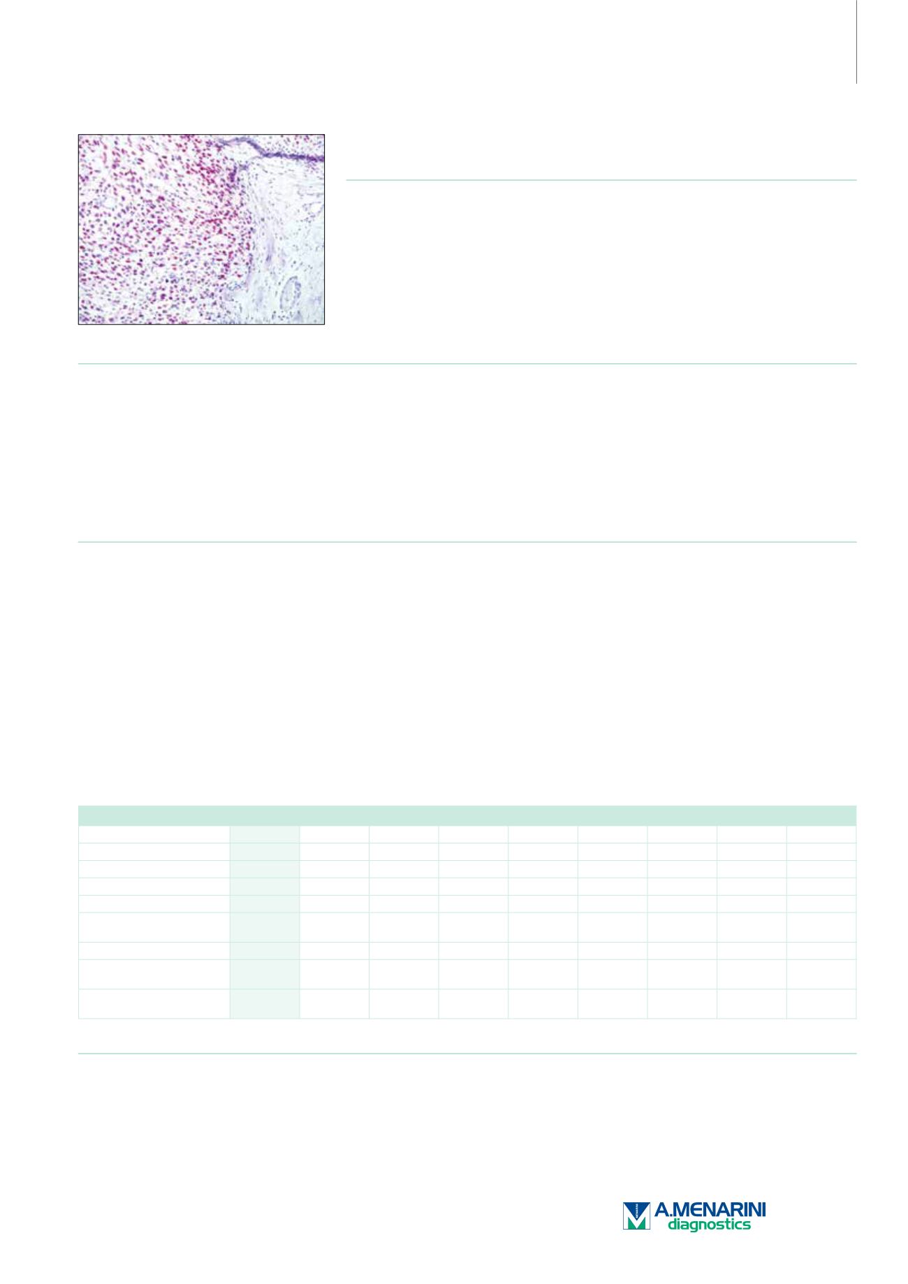
MyoD1 (EP212
†
)
Rabbit Monoclonal Antibody
Cat. No. Description
Volume
45638 MyoD1 RTU R (EP212)
7 ml Ready To Use
45620 MyoD1 0,1 R (EP212)
100 µl liquid Concentrated
45621 MyoD1 1 R (EP212)
1 ml liquid Concentrated
Product Specifications
Designation
IVD
Reactivity
Paraffin
Visualization
Nuclear
Control
Rhabdomyosarcoma
Stability
Up to 36 mo. at 2-8°C
Isotype
IgG
Manual Protocol*
• Pretreatment: Heat Induced Epitope
Retrieval (HIER)
• Primary Antibody Incubation Time:
10-30min @ 25-37°C
• 2-step polymer detection
*Please refer to product insert for complete protocol.
Product Description
Rhabdomyosarcomas (RMS) are the most frequent malignant soft tissue tumors of childhood. The spectrum of histological differentiation of this
tumor varies, as better differentiated RMS have cross-striations or rhabdomyoblasts that allow for a confident morphologic diagnosis without
adjunct studies. However, less differentiated RMS resemble other small blue round cell tumors. In at least 20% of RMS cases, IHC has been
reported to be a valuable tool in helping to make the definitive diagnosis. MyoD1, one of the MyoD family of myogenic helix-loop-helix transcription
factors, combined with myogenin, plays a role in coordinating the myogenic differentiation pathway from the determination of mesodermal
precursors into myoblasts, the differentiation of myoblasts into myotubes, and finally the maturation of myotubes into skeletal myofibers. MyoD1
is expressed in myoblasts before differentiation while myogenin has post-differentiation functions. The anti-MyoD1 immunostain identifies cells
committed to myogenesis in their earliest phase. Anti-MyoD1 has shown high sensitivity and specificity for RMS. Only intranuclear staining
should be considered as positive, since occasionally this antibody can produce a cytoplasmic staining pattern. Anti-MyoD1 could also be useful
in separating alveolar RMS from embryonal RMS. Anti-MyoD1 should be used along with anti-myogenin and anti-desmin as a trio of markers
since RMS is virtually never negative for all three of these markers. MyoD1 also stains the rhabdomyoblastic component in pleuropulmonary
blastomas and germ cell tumors, as well as reactive/regenerative myocytes in patients with other lesions such as post-chemotherapy tissue or
ulcer bed in which residual tumor was not identified.
Neoplasms
MyoD1 Myogenin Desmin
MSA
SMA
GATA3
PAX-8 CK Cocktail
SOX-10
Rhabdomyosarcoma
+
+
+
+
-
-
-
-
-
Leiomyosarcoma
-
-
+
+
+
-
-
-
-
Epithelioid Sarcoma
-
-
-
-
-
-
-
+
-
Carcinosarcoma
-
-
-
-
-
-
-
+
-
Malignant Peripheral Nerve
Sheath Tumor
-
-
-
-
-
-
-
-
+
Ewing's Sarcoma
-
-
+/-
-
-
-
-
-/+
-
Sarcomatoid Urothelial
Carcinoma
-
-
-
-
-
+/-
-
+/-
-
Sarcomatoid Renal Cell
Carcinoma
-
-
-
-
-
-
+
+/-
-
Reference
1. Morotti RA, et al. Am J SurgPathol. 2006; 30:962-968.
2. Sebire NJ, et al. J. Clin. Pathol. 2003; 56:412-416.
175


