Antibodies for
Immunohistochemistry
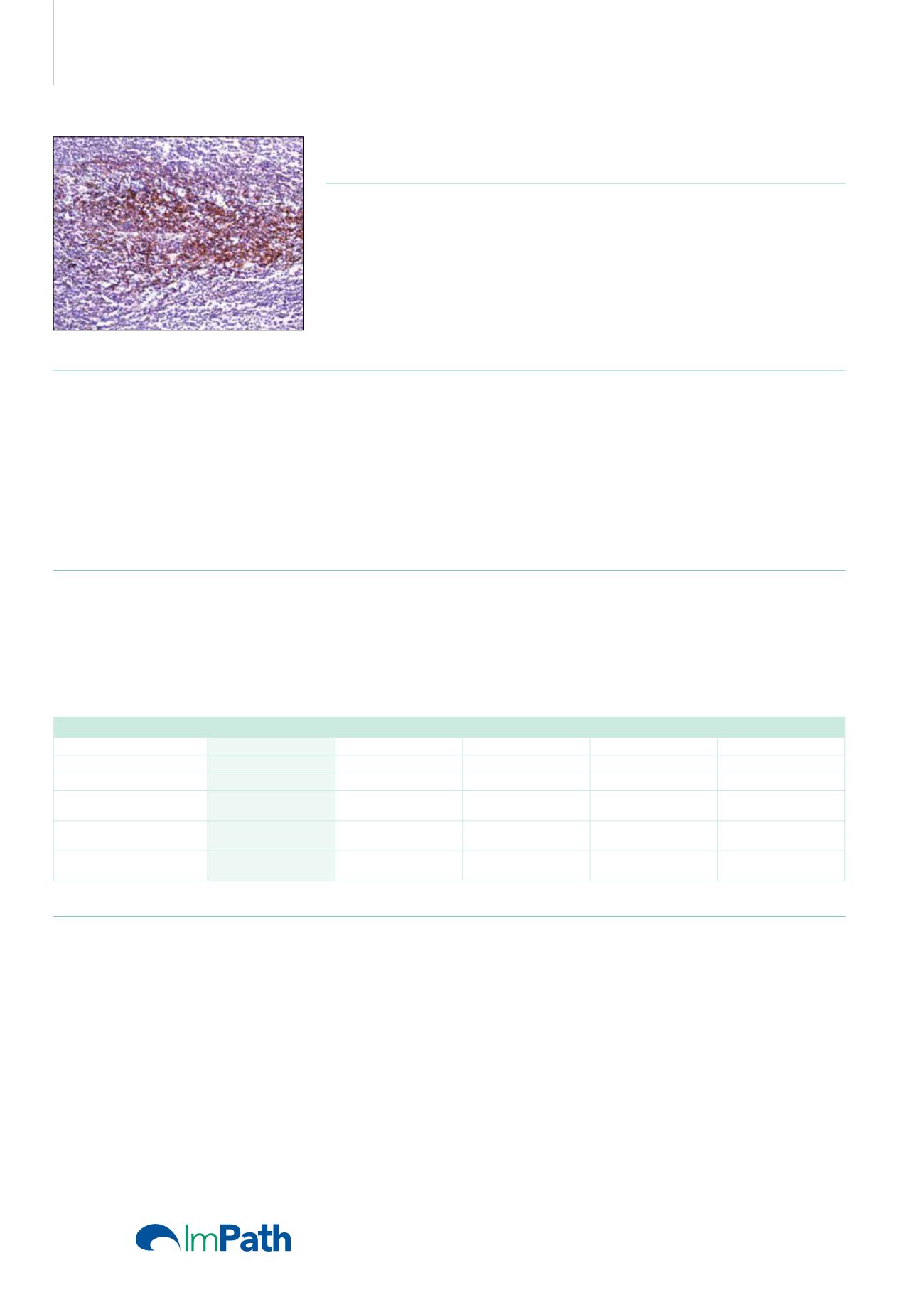
CD35 (RLB25)
Mouse Monoclonal Antibody
Cat. No. Description
Volume
45182 IMPATH CD35 RTU M (RLB25)
50 Tests
44236 CD35 RTU M (RLB25)
7 ml Ready To Use
44494 CD35 0,1 M (RLB25)
100 µl liquid Concentrated
44495 CD35 1 M (RLB25)
1 ml liquid Concentrated
Product Specifications
Designation
IVD
Reactivity
Paraffin
Visualization
Membranous
Control
tonsil
Stability
Up to 36 mo. at 2-8°C
Isotype
IgG
2b
Manual Protocol*
• Pretreatment: Heat Induced Epitope
Retrieval (HIER)
• Primary Antibody Incubation Time:
10-30min @ 25-37°C
• 2-step polymer detection
*Please refer to product insert for complete protocol.
ImPath Protocol*
• Dewax: Dewax Solution 2 (DS2)
• Pretreatment: Retrieval Solution pH 9.0
(TR1) 32min @ 98-103°C
• Primary Antibody Incubation Time:
10-90min @ 25-37°C
• HRP Polymer (Universal) or AP Polymer
(Universal) for 12 min
*Please refer to product insert for complete protocol.
Product Description
CD35, complement receptor 1, is a cell membrane-bound, monomeric glycoprotein on numerous cell types including erythrocytes, leukocytes,
glomerular podocytes, and follicular dendritic cells. The primary function of CD35 is to serve as the cellular receptor for C3b and C4b, the most
important components of the complement system leading to clearance of foreign macromolecules. The Knops blood group system is a system
of antigens located on this protein. The protein mediates cellular binding to particles and immune complexes that have activated complement.
Follicular dendritic cells (FDC) are restricted to the B-cell regions of secondary lymphoid follicles. They are CD21+/CD35+/CD1a-. Anti-CD35
labels follicular dendritic cells and follicular dendritic cell sarcoma.
Lymph Node
CD21/CD35
CD68
S-100
CD1a
Lysozyme
Reactive Histiocytosis
-
+
-
-
+
Langerhans Histiocytosis
-
+
+
+
+
Sinus Histiocytosis with
Massive Lymphadenopathy
-
+
+
-
+
Follicular Dendritic Cell
Sarcoma
+
-
-
+/-
-
Dermatopathic
Lymphadenitis
-
-
+
+
+
Reference
1. Dillon KM, et al. J Clin Pathol. 2002 Oct; 55(10):791-4.
2. Pileri SA, et al. Histopathology. 2002; 41:1-29.
3. Kunihiko Maeda, et al. J Histochem Cytochem. 2002; 50:1475-1485.
4. Chan AC, et al. histopathology. 2001 Jun; 38(6):510-8.
5. Biddle DA, et al. Modern Pathology. 2002; 15:50-58.
6. Cheuk W, et al. Am J Surg Pathol. 2001 Jun; 25(6):721-31.
7. Chang KC, et al. J Pathol. 2003 Nov; 201(3):404-12.
66


