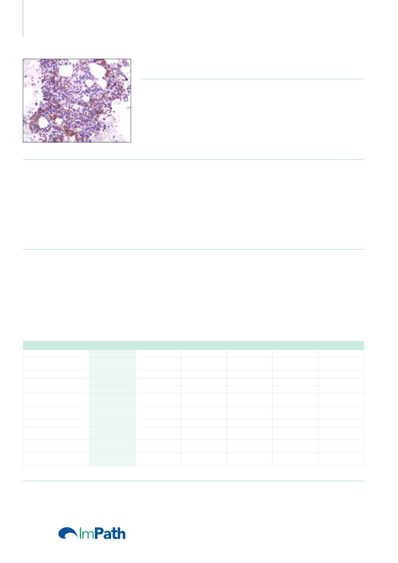
Antibodies for
Immunohistochemistry
CD33 (PWS44)
Mouse Monoclonal Antibody
Cat. No. Description
Volume
45130 IMPATH CD33 RTU M (PWS44)
50 Tests
44234 CD33 RTU M (PWS44)
7 ml Ready To Use
44490 CD33 0,1 M (PWS44)
100 µl liquid Concentrated
44491 CD33 1 M (PWS44)
1 ml liquid Concentrated
Product Specifications
Designation
IVD
Reactivity
Paraffin
Visualization
Membranous
Control
Acute myeloid leukemia with
monocytic differentiation or with
minimal differentiation, Placenta
syncytiotrophoblasts
Stability
Up to 36 mo. at 2-8°C
Isotype
IgG
2b
Manual Protocol*
• Pretreatment: Heat Induced Epitope
Retrieval (HIER)
• Primary Antibody Incubation Time:
10-30min @ 25-37°C
• 2-step polymer detection
*Please refer to product insert for complete protocol.
ImPath Protocol*
• Dewax: Dewax Solution 2 (DS2)
• Pretreatment: Retrieval Solution pH 9.0
(TR1) 32min @ 98-103°C
• Primary Antibody Incubation Time:
10-90min @ 25-37°C
• HRP Polymer (Universal) for AP 2-step
Polymer (Universal) 12 min
*Please refer to product insert for complete protocol.
Product Description
CD33 (gp67, or siglec-3) is a 67 kDa glycosylated transmembrane protein that is a member of the sialic acid–binding immunoglobulin-like
lectin (siglec) family. The genomic locus of this protein has been mapped to chromosome 19q13.1-3.5. In maturing granulocytic cells, there is
progressive down-regulation of CD33 from the blast stage to mature neutrophils. However, in monocytes and macrophages/histiocytes, strong
expression of CD33 is maintained throughout maturation. Therefore, the positive control tissue should be bone marrow myeloid cells (especially
myeloid precursors), liver Kupffer cells, lung alveolar macrophages, or placental syncytiotrophoblasts. Detection of CD33 using monoclonal
antibodies has been a critical component in immunophenotyping acute leukemias, particularly acute myeloid leukemias. This anti-CD33 may
be particularly advantageous for cases of acute myeloid leukemia, minimally differentiated (AML-M0) and acute monocytic leukemia (AML-M5),
in which other paraffin section markers of myeloid differentiation (such as anti-myeloperoxidase) may be negative. However, anti-CD33 staining
cannot be used in isolation and must be correlated with other myeloid and lymphoid markers because cases of myeloid antigen–positive acute
lymphoblastic leukemia may show bona fide CD33 expression.
Neoplasms
CD33
CD34
CD117
CD71
CD163
MPO
AML with Minimal
Differentiation
+
+
+
-
-
-/+
AML without Differentiation
+
+
+
-
-
-/+
AML with Maturation
+
+
+
-
-
+
APL
+
-
+
-
-
+
Acute Myelomonocytic
Leukemia
+
+/-
+/-
-
+
+/-
Acute Monoblastic and
Monocytic Leukemia
+
+/-
+/-
-
+
-/+
Acute Erythroid Leukemia
+
-
+/-
+
-
-
Acute Megakaryoblastic
Leukemia
+/-
-
-
-
-
-
B-lymphoblastic Leukemia/
Lymphoma
-/+
+/-
-
-
-
-
T-lymphoblastic Leukemia/
Lymphoma
+/-
+/-
-
-
-
-
Reference
1. Crocker PR, et al. Biochem Soc Symp. 2002; 69:83-96.
2. Braylan RC, et al. Cytometry. 2001; 46:23-27.
3. Chang H, et al. Leuk Res. 2004; 28:43-48.
64


