Advanced Solutions
for Advanced Pathology
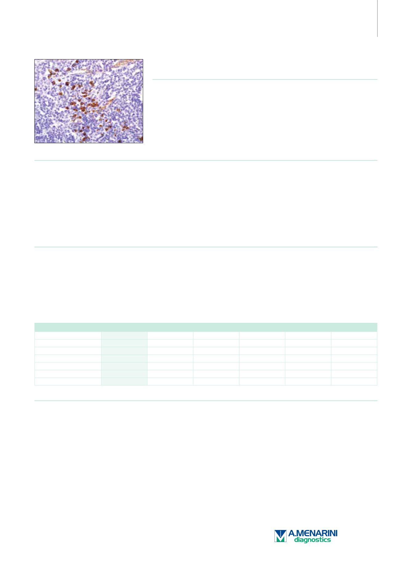
IgA (Polyclonal)
Rabbit Polyclonal Antibody
Cat. No. Description
Volume
45217 IMPATH IgA RTU R (Poly)
50 Tests
44317 IgA RTU R (Poly)
7 ml Ready To Use
44654 IgA 0,1 R (Poly)
100 µl liquid Concentrated
44655 IgA 1 R (Poly)
1 ml liquid Concentrated
Product Specifications
Designation
IVD
Reactivity
Paraffin
Visualization
Cytoplasmic
Control
Tonsil
Stability
Up to 36 mo. at 2-8°C
Manual Protocol*
• Pretreatment: Heat Induced Epitope
Retrieval (HIER)
• Primary Antibody Incubation Time:
10-30min @ 25-37°C
• 2-step polymer detection
*Please refer to product insert for complete protocol.
ImPath Protocol*
• Dewax: Dewax Solution 2 (DS2)
• Pretreatment: Retrieval Solution pH 9.0
(TR1) 32min @ 98-103°C
• Primary Antibody Incubation Time:
10-90min @ 25-37°C
• HRP Polymer (Universal) or AP Polymer
(Universal) for 12 min
*Please refer to product insert for complete protocol.
Product Description
Immunoglobulin A (IgA) is a class of antibodies which plays a critical role in mucosal immunity. More IgA is produced in mucosal linings than all other
typesofantibodycombined;between3gand5g issecreted intothe intestinal lumeneachday.In itssecretoryform, it isthemain immunoglobulinfound
in mucous secretions, including tears, saliva, colostrum, intestinal juice, vaginal fluid, and secretions from the prostate and respiratory epithelium.
It is also found in small amounts in blood. Because it is resistant to degradation by enzymes, secretory IgA can survive in harsh environments such as
the digestive and respiratory tracts, to provide protection against microbes that multiply in body secretions. IgA is a poor activator of the complement
system, and opsonizes only weakly. Its heavy chains are of the type α. IgA exists in two isotypes, IgA1 (90%) and IgA2 (10%). IgA1 is found in serum
and ismadeby bonemarrowB-cells. IgA2 ismadebyB-cells located in themucosa andhas been found in colostrum,maternal milk, tears, and saliva.
Anti-IgA antibody reacts with surface immunoglobulin IgA alpha chains. It is useful when identifying plasma cell myeloma.
Immunoglobulin, Heavy and Light Chain
IgA
IgG
IgD
IgM
Kappa
Lambda
Secretory Meningioma
+
-
-
+
Cutaneous Lymphoma
-
-
-
-
+/-
-/+
Myeloma
+
+
-/+
-/+
+/-
-/+
Diffuse LBCL
-
+
-
+
+/-
-/+
Marginal Zone Lymphoma
-
-
-/+
+
+/-
-/+
SLL/CLL
-
-
+
+
+/-
-/+
Reference
1. Arnold A, et al. New Eng J Med. 1983; 309:1593-1599
2. Leong AS, et al. Manual of Diagnostic Antibodies for Immunohistology. Geenwich Medical Media Ltd. 1999. pp 217-219.
3. Hertel BF, et al. New Eng J Med. 1980; 302:1293-1297.
4. Taylor CR. Arch Path Lab Med. 1978; 102:113-121.
5. Warnake R, et al. Masson Publishing USA. 1981. pp 203-221.
145


